 iSmartNetwork Magazine The online magazine for you…
Testing cancer treatments is a lot of trial and error currently, and patients are often subject to multiple uncomfortable and time-consuming therapies before obtaining one that the project works. Progress have been induced, including growing artificial tumors to test medicines on specific cancer sorts, but these tumors can take weeks to grow and they don’t account for patients’ individual biological makeup. Now, however, researchers from MIT and Draper Universityhave come up with a brand-new option: a 3D published microfluidic machine that simulates cancer treatments on biopsied cancerous tissue.
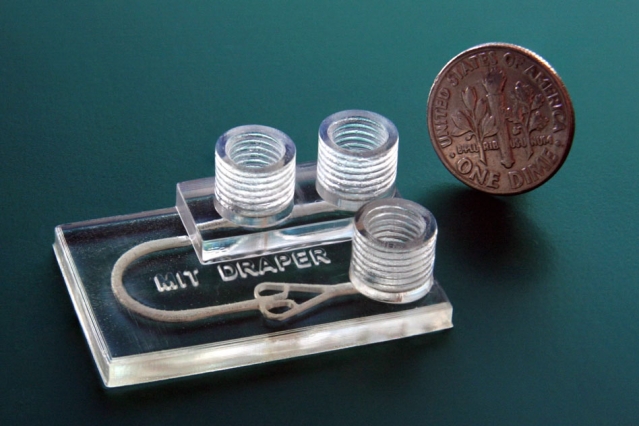
The device is a chip slightly larger than a one-quarter that can be 3D printed in about an hour. It has three cylindrical chimneys protruding from the surface, who the hell is ports that input and drain fluids as well as remove unwanted breath froths. The biopsied tumor fragments are placed in a chamber attached to a system of deliver fluids to the tissue. These fluids could contain things like immunotherapy agents or immune cells. Clinicians can then use imaging techniques to see how the tissue responds to the treatments.
The researchers use a new type of biocompatible resin, traditionally to be applied for dental applications, that can support the long-term survival of biopsied tissue. This compares with other 3D printed microfluidic medicine experimenting machines, which have substances in the resin that speedily kills the cells. Fluorescence microscopy images showed that the new device, called a tumor analysis platform or TAP, preserved more than 90 percentage of the tissue alive for at the least 72 hours and potentially much longer.
The TAP is cheap and easy to fabricate, so it could promptly be implemented into clinical puts, according to the researchers. The machines is adaptable as well- physicians could 3D print a multiplexed device who are able to substantiate multiple tumor samples in similarity, so that the interactions between tumor fragments and several different stimulants could be modeled simultaneously for a single patient.
” People anywhere in the world could print our designing. You can foresee a future where your doctor will have a 3-D printer and can print out the machines as needed ,” said Luis Fernando Velasquez-Garcia, a researcher in the Microsystems Technology Laboratory.” If someone has cancer, you can take a bit of tissue in our machine, and keep the tumor alive, to run multiple tests in latitude and figure out what would work best with the patient’s biological makeup. And then enforce that treatment in the patient .”
One potential application is experimenting immunotherapy, a brand-new therapy technique that uses narcotics to “rev up” a patient’s immune system to help it fighting cancer.
” Immunotherapy treatments have been specifically developed to target molecular markers found on the surface of cancer cells ,” said alumnu researcher Ashley Beckwith.” This helps to ensure that the care elicits an attack on the cancer directly while limiting negative impacts on health tissue. Nonetheless, every individual’s cancer shows a unique array of surface molecules — as such, it can be difficult to predict who will respond to which therapy. Our device uses the actual tissue of the person, so is a perfect fit for immunotherapy .”
The research was published in a paper entitled” Monolithic, 3D-Printed Microfluidic Platform for Recapitulation of Dynamic Tumor Microenvironments .”
 ” A key challenge in cancer experiment has been the process of developing tumor microenvironments that simulate mechanisms of cancer advance and the tumor-killing the consequences of novel therapeutics ,” said Jeffrey T. Borenstein, who leads the immuno-oncology program at Draper.” Through this collaboration with Luis and the MTL, we are able to benefit from their great expertise in additive manufacturing technologies and materials science for extremely rapid designing cycles in house and testing these systems .”
Microfluidic devices are normally grew via micromolding with PDMS. The technique was not suitable, nonetheless, for producing a device with fine 3D aspects such as the fluid canals, so the researchers turned to 3D printing, which allowed them to create the machine in one piece. They experimented with several resins, but ultimately settled on Pro3dure GR-1 0, which is often used to induce mouth guards. The resin is nearly as transparent as glass, can be printed in very high resolution, and has hardly any surface imperfections- and it doesn’t is harmful to cells.
” When you publish some of these other resin substances, they radiate chemicals that mess with cells and kill them. But this doesn’t do that ,” Velasquez-Garcia said.” To the best of my knowledge, there’s no other printable material that comes close to this degree of inertness. It’s as if the material isn’t there .”
The device also features a” bubble trap” and a” tumor trap .” Fluids flowing into a device like this one creates foams that can disrupt the experiment or flare and liberate breath that destroys tumor tissue. So health researchers developed a bubble trap, a chimney that rises from the liquid canal into a threaded port through which air flees. Fluid get inserted into an inlet port adjacent to the net, then flows past the net, where any bubbles in the liquid rise up through the threaded port and out of the device. Fluid is then routed around a small U-turn into the tumor’s chamber, where it pours through and all over the tumor fragment.
The tumor trap sits at the intersection of the larger inlet channel and four smaller inlet channels. Tumor fragments, less than one millimeter across, are administered into the inlet canal via the bubble trap. As the fluid flows through the device, the tumor is steered downstream to the tumor trap, where it gets caught. The fluid continues traveling along the outlet canals, which are too small for the tumor to fit into, and drains out of the machine. A continuous pour of fluids keeps the tumor scrap in place and forever replenishes nutrients for the cells.
” Because our device is 3-D printed, we were able to establish the geometries we wanted, in the materials we wanted, to achieve the performance we wanted, instead of compromising between what was designed and what could be implemented — which typically happens when using standard microfabrication ,” Velasquez-Garcia said.
The next step is to experiment how the tumor fragments respond to therapeutics.
” The traditional PDMS can’t make such structures you need for this in vitro environment that can retain tumor fragments alive for a significant period of time, ” said Roger Howe, a prof of electrical engineering at Stanford University, who was not involved in the research.” That you can now induce quite complex fluidic enclosures that will allow most realistic contexts for experimenting out various narcotics on tumors speedily, and potentially in clinical lays, is a major contribution .”
Discuss this and other 3D publication topics at 3DPrintBoard. com or share your thoughts below.
[Source/Images: MIT]
Read more: 3dprint.com